Li-Ying Fan1,2,
Te Guo1,
Jun Ren3,
Yi-Fa Zhou1 ![]()
For correspondence:- Yi-Fa Zhou Email: Zhouyf383@nenu.edu.cn Tel:+8643185098212
Received: 2 March 2016 Accepted: 5 October 2016 Published: 29 November 2016
Citation: Fan L, Guo T, Ren J, Zhou Y. Effect of ginseng root polysaccharides on cutaneous wound repair in mice. Trop J Pharm Res 2016; 15(11):2399-2405 doi: 10.4314/tjpr.v15i11.14
© 2016 The authors.
This is an Open Access article that uses a funding model which does not charge readers or their institutions for access and distributed under the terms of the Creative Commons Attribution License (http://creativecommons.org/licenses/by/4.0) and the Budapest Open Access Initiative (http://www.budapestopenaccessinitiative.org/read), which permit unrestricted use, distribution, and reproduction in any medium, provided the original work is properly credited..
Purpose: To evaluate the healing activity of water ginseng (Panax ginseng C.A. Meyer) polysaccharide (WGP) in vivo.
Methods: Mice were divided into four groups: group 1 was the control, and groups 2–4 were treated with WGP (15 mg or 30 mg) or mouse epidermal growth factor (mEGF, 0.1 mL, 2000 IU/mL), respectively. Full-thickness excision wounds were made on the back of each mouse. Wound areas were imaged, and wound contraction (%) was calculated. Histological observation and analysis of collagen content were performed using standard biochemical methods.
Results: Wound contraction and the period of epithelialization in WGP groups were significantly different from those of control. The 30 mg WGP group showed 85.0 % wound healing, while control group showed only 65.8 % wound healing (p < 0.001) 10 days after the operation. WGP also promoted the formation of collagen in a dose-dependent manner. Histological examination confirmed that fibroblasts and new blood vessels were more abundant in WGP group than in control group.
Conclusion: Topical application of WGP stimulates wound contraction and accelerates healing. This may be due to the ability of WGP to enhance the functions of macrophages and myeloid hematopoietic cells, resulting in increased fibroblast division, collagen synthesis, and production of blood cells during wound healing.
Introduction
The skin plays a crucial role in maintaining homeostasis. Wounds are breaks in the epithelial integrity of the skin, and may result in disruption of the structure and function of the underlying normal tissue, as well as other complications, including bacterial infections and fluid loss. Therefore, continuity of the skin must be expeditiously restored [1]. Wound healing involves a dynamic, well-orchestrated series of events that involves many different types of cells and inflammatory mediators such as cytokines [2,3]. Macrophages are essential for the transition from the inflammation to repair phases, and help orchestrate fibroblast proliferation and the removal of fibrin. Macrophages also secrete tumor necrosis factors (TNFs), which stimulate both fibroblasts and keratinocytes [4]. Fibroblasts proliferate and produce matrix proteins, including collagen, which help construct a new extracellular matrix that supports further ingrowths of cells and is essential for the repair process. Wound contraction occurs via an interaction between fibroblasts and the surrounding extracellular matrix [3].
Polysaccharides from plant extracts are important macromolecules that have been used as therapeutic agents in both animals and humans. In traditional medicine, polysaccharide-containing plant extracts are widely employed for the treatment of skin and epithelium wounds and mucous membrane irritation [5]. Ginseng has been used for more than 2000 years, and is generally considered a longevity medicine [6]. Chinese traditional medicine textbooks describe its ability to tonify blood, expel toxins, promote skin regeneration, prevent infections, and regulate the immune system, among other actions [7–11]. Ginseng extracts have also been used clinically as a topical treatment for atopic suppurative dermatitis, wounds, and skin inflammation. Ginseng polysaccharides are major active components of ginseng [12,13]. Numerous studies on the pharmacology of WGP have shown that they induce the secretion of TNFs, enhance the activity of T cells [12,14], increase the function of mononuclear phagocytes, and promote the transformation of mononuclear phagocytes into macrophages [15]. WGP also play an important role in regulating the proliferation and differentiation of myeloid hematopoietic cells, which may be related to increases in hepatocyte growth factor (HGF) secreted by stromal cells in hematopoietic microenvironments to promote the production of blood cells [16,17]. However, studies on the effects of WGP on wound repair are lacking.
Therefore, we investigated the effects of WGP on the healing of full-thickness skin wounds in mice, as well as collagen deposition and the mechanisms by which WGP promote healing.
Methods
Materials
WGP was prepared according to a previously reported method [18]. Dry ginseng root was heated in distilled water at 100 °C for 3 – 4 h. The extract filtrate was condensed in a rotary evaporator and mixed with cold absolute ethanol. The polysaccharide precipitates were collected from the alcohol phase by centrifugation (4500 rpm, 10 min). To obtain a pure product, the polysaccharides were deproteinized using Sevag reagent (n-butanol:chloroform = 1:4), starches were removed using α-amylase, and the polysaccharides were precipitated with 75 % ethanol. After dialysis using distilled water, followed by lyophilization, WGP was obtained consisting of 84.4 % (w/w) sugar and 34.6 % (w/w) uronic acid. The monosaccharide component consisted of 41.2 % galacturonic acid, 20.1 % galactose, 16.1 % glucose, 15.8 % arabinose, 2.7 % rhamnose, and 2.2 % mannose. Before using them, they were prepared as gels by dissolving 15 mg or 30 mg lyophilized powder in 0.1 mL distilled water. mEGF was purchased from Sigma Co., USA. Paraffin was kindly provided by Leica. Co. Ltd., Germany. A hydroxyproline assay kit was purchased from Sigma Co., USA. Slide glass was supplied by Matsunami, Japan. All chemicals and solvents were of analytical grade.
Animals
Forty male and female ICR mice weighing 28–32 g were provided by the Animal Center of Jilin University. Animals were acclimatized for a week in a room with a controlled temperature of 23 ± 2 °C and a relative humidity of 30–60 %, and given free access to standard laboratory diet and water prior to the investigation. All animal experiments were approved by the Animal Experimental Ethics Committee of Northeast Normal University (approval ref no. 20130110) and carried out in strict accordance with the recommendations in the Guide for the Care and Use of Laboratory Animals of the National Institutes of Health [19].
Excision wound model
The mice were divided into four groups of forty animals: group 1 was the control, and groups 2–4 were treated with WGP (15 mg), WGP (30 mg), or mEGF (0.1 mL, 2000 IU/mL), respectively. The animals were anesthetized with ether, the dorsal hair was clipped, and one full-thickness, round wound about 9 mm in diameter was created on the back of each mouse using a sharp pair of scissors and a scalpel. Then WGP was applied to the wounds at a dose of 15 mg or 30 mg. The control wound was treated with sterile cotton gauze during the postoperative period. After surgery, the animals were kept individually in separate cages. The progressive changes in wound area were measured in mm2 by tracing the wound boundaries on a plastic sheet every other day. Tracings were digitized by scanning and wound areas were quantified using Image J software. Wound contraction was expressed as the percentage reduction in wound size, as formulated in Eq 1.
PW = {(S0– SN)/S0} × 100 ……………………. (1)
where PW is the percentage of wound contraction, S0 (mm2) is the primary wound area, and SN (mm2) is the wound area on day N.
Biochemical assays
Collagen is an important component of wound healing, as it fills in tissue defects. Hydroxyproline exists almost exclusively in collagen [18] and thus is a sensitive biochemical marker that reflects changes in collagen fibers. Concentrations of hydroxyproline and collagen were estimated using the traditional acid hydrolysis method [20,21]. Excised granulation tissues were weighed, defatted in a chloroform:methanol mixture (2:1, v:v), hydrolyzed in 6.0 N HCl for 6 h at 110 °C, and evaporated to dryness. The hydrolysate was neutralized to pH 6.0–6.8 and subjected to chloramine-T oxidation. Then the test tubes were placed in a water bath at 60 °C for 25 min. The reaction was terminated by adding 1 mL (0.4 M) perchloric acid, the color was developed by adding 1 mL paradimethyl aminobenzaldehyde (PDAB), and the assay was read at 557 nm using a spectrophotometer. The collagen content was estimated from the hydroxyproline concentration by multiplying by 7.51 [20].
Histological examination
Skin including the wound was carefully excised 1, 3, 7, and 10 days after injury and fixed in 4 % formaldehyde, embedded in paraffin, and cut in 4–6 μm increments with a microtome. The sections were made perpendicular to the anterior–posterior axis and perpendicular to the surface of the wound. The sections were positioned on a slide, and stained with hematoxylin-eosin (HE) reagent. Representative sections from each wound were quantitatively evaluated for wound width, wound cellularity, neovascularization, and tissue granulation.
Statistical analysis
All data are expressed as mean ± SD. Statistical analyses were performed using one-way ANOVA (SPSS Statistics 17.0). Differences were considered significant when p < 0.05.
Results
In vivo observation
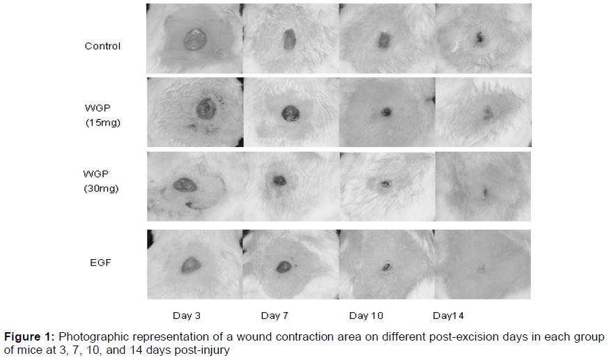
There was no mortality in the study. In vivo observations were limited to injured lesions, body weight, and diet. The lesions resulted in local redness and swelling and moist fluid exudation from the subcutaneous tissues and wound edges in both the control and study groups. Three days after surgery, wounds treated with WGP/EGF had no effusion and new granulation had begun to cover part of the wound, while the control wounds still had some effusion and no granulation (). WGP/EGF treatment considerably decreased inflammatory cells in the early phase of healing. There were no differences in body weight or diet between the control and study groups.
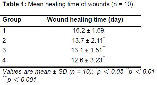
Healing time, wound area, and wound contraction
To evaluate the effects of WGP on wound healing, healing time and closure were observed. Healing time was significantly slower in controls than in the treatment groups (). WGP treatment accelerated wound contraction and shortened the healing time by 2–3 days. In addition, by day 7, wounds treated with 30 mg WGP had more epithelial tissue than the controls ().
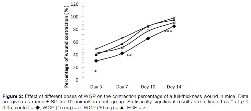
As shown in , treatment of wounds with 15 mg and 30 mg WGP resulted in a higher rate of wound contraction, and the 30 mg WGP group showed a faster closing rate. The rate of wound contraction in control mice was 30.2 to 42.2 % from day 3 to day 7 and 65.8 to 85.3 % from day 10 to day 14; complete epithelialization and healing were observed on day 16. Mice treated with 30 mg WGP showed increased wound contraction from 40.5 % on day 3 to 57.4 % on day 7 and 85.0 to 95.0 % from day 10 to day 14. Complete epithelialization and healing were observed on day 13. The percent of wound contraction in mice treated with mEGF was 49.3 % on day 3, 66.4 % on day 7, and 85.1 % on day 10 to 98.3 % on day 14, which was similar to the 30 mg WGP group.
Biochemical profile
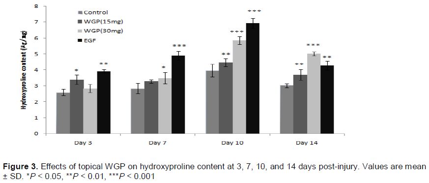
presents changes in collagen content. The maximum concentration of collagen in wounds from all groups was observed on day 10 post-injury. Both concentrations of WGP resulted in a significantly higher level of collagen compared to controls at all time points (p < 0.01). Then the concentration gradually decreased from day 10 and returned to the level recorded on day 7 in all experimental groups. However, the wounds from animals in groups 2 and 3, which were treated with WGP, had a higher level of collagen than those in group 1 throughout the treatment (p < 0.05, ).
Histological features
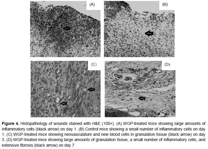
Histological examination of wounds supported the observation that WGP accelerated healing. A significant increase in inflammatory cell infiltration and macrophage phagocytosis was found in WGP-treated wounds compared to control wounds after 1 day (A,B). Skin wounds treated with WGP showed better healing than control mice, including observable granulation and the formation of a stratified epithelium (D). Furthermore, neovasculature in granulation tissue progressed faster in WGP-treated mice compared to control mice (C). By day 10 after injury, WGP-treated wounds were almost completely epithelialized and almost all necrotic tissues had been replaced by new granulation. The wounds were well protected and firmly covered with scabs.
Discussion
We evaluated the wound-healing potential of WGP applied to full-thickness wounds in mice. Numerous studies have shown that the topical application of WGP can stimulate wound contraction and improve healing [23,26–28]. We found that the application of 15 mg or 30 mg WGP induced significant wound healing from days 3 to 14 after injury compared to the control group. Wound closure in the 30 mg WGP group was similar to that in the EGF-treated group, and healing was significantly higher than in controls (40.5 vs. 30.2 %, 57.4 vs. 42.2 %, 85.0 vs. 65.8 %, and 95.0 vs. 85.3 % on days 3, 7, 10, and 14, respectively). The maximum difference between the WGP groups and controls was recorded 10 days after injury (). Healing time was also shorter in WGP-treated mice than in controls. Together, these results suggest that WGP can accelerate wound contraction, and that higher doses have a stronger effect.
The healing process begins with cell proliferation. The main cells that trigger the process are macrophages that remove foreign bodies and direct the development of granulation tissue. Then fibroblasts and endothelial cells migrate towards the injured area, increasing tissue permeability and the production of collagen fiber [22]. Wound contraction occurs through the interaction between fibroblasts and the surrounding extracellular matrix. Histological examinations confirmed that fibroblasts were abundant 3 days after injury in the WGP-treated groups. During the phase in which fibroblasts are attracted to the wounds, angiogenesis occurs from blood vessels near the injury. In WGP-treated groups, new blood vessels and soft, pink granulation tissue were well established. In contrast, in the control group, fibroblast cell proliferation was slow and new blood vessels and granulation tissue were not obvious 3 days after injury. These effects may be due to the increase in HGF secretion from stromal cells induced by WGPs in the hematopoietic microenvironment, which promotes the development of blood cells [13]. HGF is a key growth factor involved in wound healing, and it plays an important role in neovascularization and the formation of granulation tissue [1]. These findings suggest that WGP accelerate wound closure in mice to a similar extent as EGF.
Three days post-injury, control wounds were obviously red and swollen, with inflammatory exudation. In contrast, WGP-treated wounds were almost completely dry and covered with granulation tissue with no obvious swelling () [2]. These results suggest that WGP can increase the function of the mononuclear phagocyte system [14], which is an important process in wound healing. In the early phases of wound healing, monocytes are recruited to the wound by growth factors, and monocyte chemo-attractant proteins adhere to the provisional fibrin plug matrix via integrin receptors before transforming into macrophages. Macrophages perform phagocytosis to destroy invading bacteria and clear debris and necrotic tissue [3]. Therefore, WGP treatment decreased the inflammatory response at the wound site (A). This effect could also be related to the antimicrobial traits of WGP [20,22].
Hydroxyproline is an amino acid that is essential for the synthesis of collagen fibers. For this reason, its levels are used as an indicator of collagen content. Hydroxyproline levels in granulation tissues increased in all treatment groups from days 3 to 10, and then returned to day 7 levels by day 14 () [24–26].
Conclusion
Application of WGP to open wounds induces significant wound contraction and accelerated closure and healing in mice. These results lend some support to the traditional use of ginseng root to treat skin diseases. However, more work is required to unravel the possible mechanisms involved in the process of wound healing, whether it is by regulating the body’s immune system or by direct action on the wound.
Declarations
Acknowledgement
References
Archives


News Updates